
Lesson 5 of 13 · about 5 minutes
Seeing
From the eye to the back of the brain.

In the 3D app, the brain flies to each part as you read, and 2 quick questions at the end help it stick. Here is the whole lesson to read.
- 1
The optic nerve
Each eye sends its picture down an optic nerve of about a million fibers.
More about the optic nerve (II) →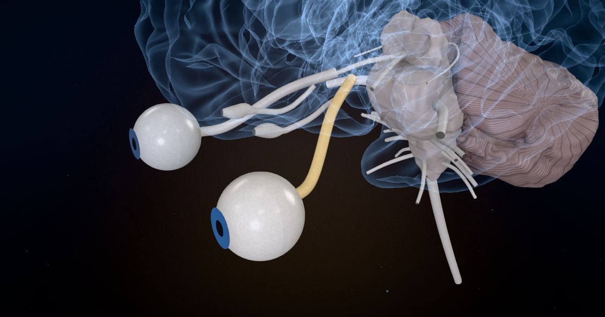
- 2
The crossing
At the optic chiasm, fibers from the inner half of each eye cross over, so each side of the brain sees the opposite half of the world.
More about the optic chiasm →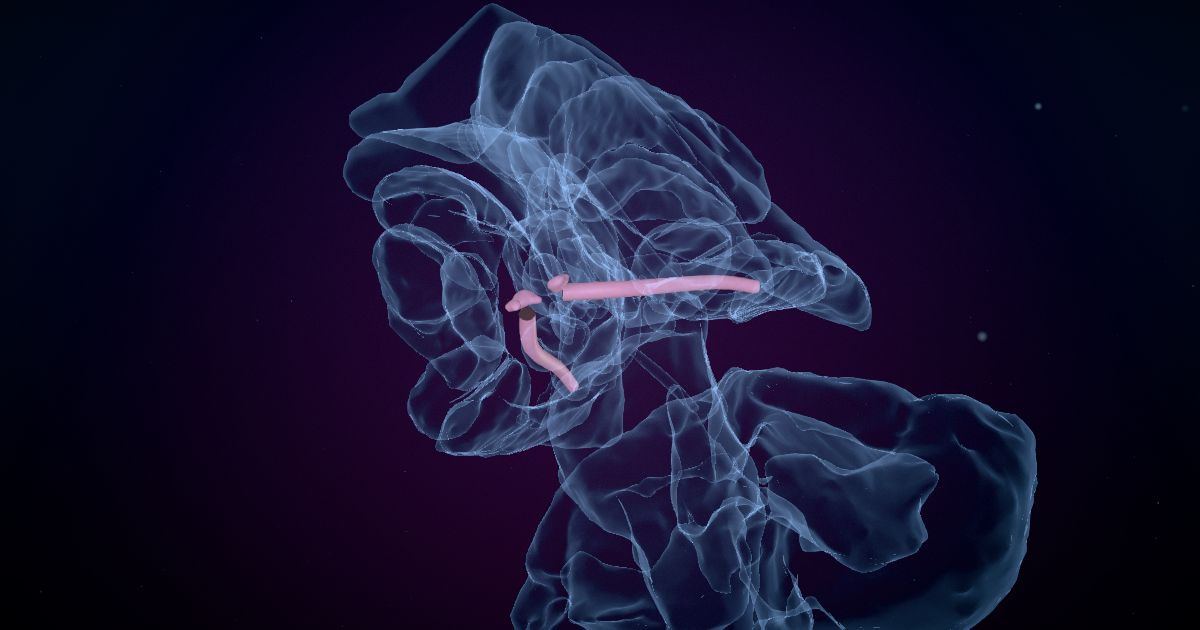
- 3
The relay
The lateral geniculate nucleus of the thalamus sorts the signals and passes them on.
More about the lateral geniculate nucleus →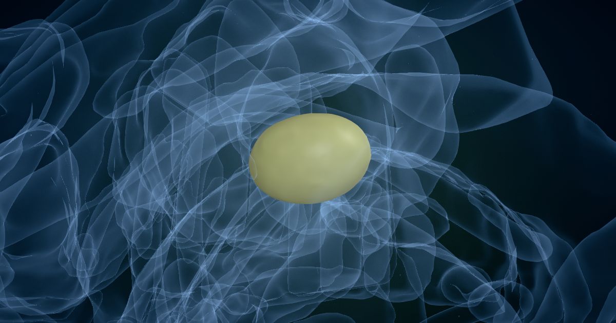
- 4
A sweeping fan
The optic radiation fans back through the white matter. Some of its fibers loop forward into the temporal lobe first.
More about the optic radiation →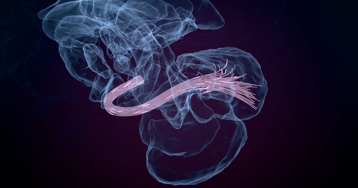
- 5
Primary visual cortex
Around the calcarine groove on the inner surface at the back, the primary visual cortex maps what you see, point by point.
More about the primary visual cortex →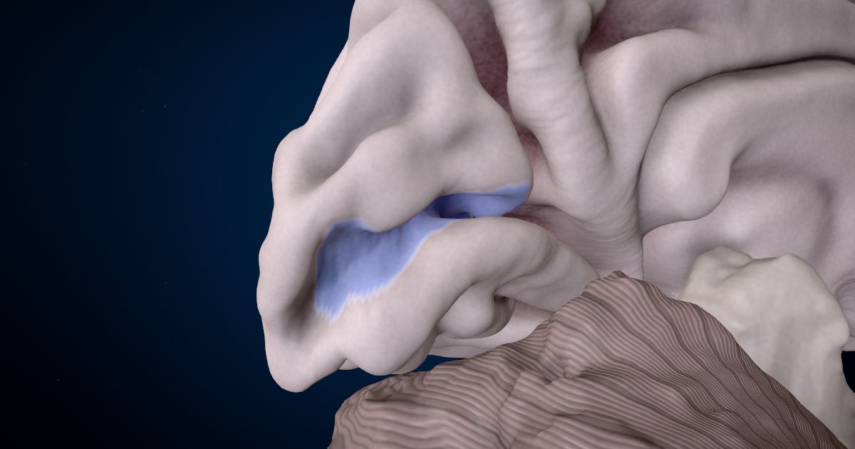
- 6
Recognizing faces
Along the underside of the temporal lobe, the fusiform gyrus recognizes faces and written words.
More about the fusiform gyrus →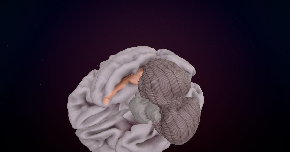


Now see it for yourself
Take this lesson in 3D: turn the brain around, look inside, and finish with 2 quick questions.
Start the lesson in 3D